Summarize This Content with Artificial Intelligence (AI):
Echocardiography (ECHO) is a type of heart ultrasound that uses sound waves to check heart and vascular health and to detect heart diseases.
What Is Echocardiography (ECHO)?
Echocardiography (ECHO) is a test that uses sound waves to visualize the function and structure of the heart. With ECHO, it is possible to observe the movements of the heart muscle, the chambers of the heart, the heart valves, and blood flow. Echocardiography does not contain radiation, does not require any incision, is painless, and is performed externally.
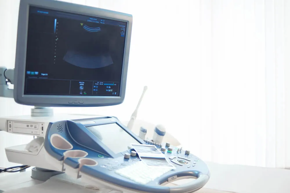
A standard echocardiography device consists of a probe and an imaging console. The echo probe emits high-frequency sound waves that are transmitted through a gel applied to the skin. The returning echoes are then displayed on the imaging screen. The imaging console includes a display monitor and a control panel that allows the operator to adjust depth, image quality, zoom, and color imaging.
Fill out the form to get information.
What Conditions Can Echocardiography Detect?
Echocardiography is one of the main imaging tests used to detect heart diseases and is commonly known as a heart ultrasound. The ultrasound imaging technique can also be applied to many other parts of the body besides the heart. Conditions that can be diagnosed with echocardiography include:
- Heart failure: Characterized by reduced pumping ability of the heart.
- Heart valve diseases: Problems such as narrowing or leakage of the heart valves.
- Pericardial diseases: Such as pericarditis or pericardial effusion.
- Congenital heart defects: Such as holes in the heart or abnormal blood vessels.
- Cardiomyopathies: Thickening or enlargement of the heart muscle.
- Pulmonary hypertension: High blood pressure in the lungs affecting the heart.
- History of heart attack: Damaged or non-moving heart muscle areas may indicate a previous heart attack.
Types of Echocardiography (ECHO)
Echocardiography is classified into different types depending on the disease and the purpose of the test. The main types include:
- Transthoracic Echocardiography (TTE)
- Transesophageal Echocardiography (TEE)
- Stress Echocardiography
- Strain and Strain Rate Echocardiography
What Is Transthoracic Echocardiography (TTE)?
Transthoracic echocardiography (TTE) is the most commonly used standard heart ultrasound. Sound waves are transmitted through a probe placed on the chest wall to visualize the structure and function of the heart. The procedure lasts about 15 minutes, is painless, radiation-free, and safe for adults, children, and pregnant women.
With TTE, the heart valves, muscle, walls, chambers, and major vessels can be visualized. The image quality may vary depending on the patient’s body type — in individuals with obesity or emphysema, imaging may take longer due to difficulty obtaining clear visuals.
What Is Transesophageal Echocardiography (TEE)?
Transesophageal echocardiography (TEE) is performed by inserting an ultrasound probe into the esophagus to obtain a closer and more detailed view of the heart. Since the esophagus lies directly behind the heart, this method provides clearer images of the heart valves, possible infections, or blood clots.
Before the procedure, a local anesthetic spray is applied to the throat, and a thin ultrasound probe is gently advanced into the esophagus. The procedure typically lasts around 30 minutes but may take longer depending on the patient’s age and condition.
What Is Stress Echocardiography?
Stress echocardiography evaluates the heart’s function under stress, either through exercise or medication. It helps detect hidden blockages in the coronary arteries. The test has two stages:
- Imaging the heart at rest.
- Imaging the heart after physical exertion or after administering medication to simulate exercise.
If an area of the heart does not receive enough blood flow during stress, its motion may appear abnormal. Stress echocardiography is also used to assess a patient’s risk of heart attack.
What Is Strain and Strain Rate Echocardiography?
Strain echocardiography precisely measures the contraction and relaxation ability of the heart muscle (myocardium). “Strain” indicates how much the heart muscle fibers deform, while “strain rate” refers to the speed of this change. This method detects early signs of heart failure or cardiomyopathy and is useful in monitoring heart damage caused by chemotherapy.
Strain and strain rate echocardiography are performed using the standard ECHO system without requiring additional equipment.
How Is Echocardiography Performed?
During a standard ECHO, the patient lies slightly on their side on an examination bed, and gel is applied to the chest. The ultrasound probe is moved across the chest at various angles and pressures to obtain detailed images of the heart. The procedure takes about 15 minutes, depending on the patient’s body type and the purpose of the examination.
What Does an Echocardiography Report Include?
The report provides detailed information about the structure and function of the heart, including the size of heart chambers, wall thicknesses, valve conditions, pericardium, and major vessels. Key parameters include chamber diameters, wall thickness, and ejection fraction (EF).
How Are Echocardiography Results Interpreted?
ECHO results are interpreted by a cardiologist. The most important measurement is the ejection fraction (EF), which shows how effectively the heart pumps blood. A normal EF value is between 50% and 70%. Lower values may indicate heart failure or weakened heart muscle.
Echocardiography results may require additional tests or blood analyses for a complete diagnosis.
Frequently Asked Questions About Echocardiography
How long does echocardiography take?
A standard ECHO takes about 15 minutes, but this can vary depending on the test type and patient.
Why is contrast echocardiography performed?
Contrast ECHO uses a special contrast agent injected intravenously to enhance image clarity when standard imaging is insufficient.
How is echocardiography performed in children?
It is performed the same way as in adults. In some cases, a mild sedative may be given. The test is painless and radiation-free.
Can echocardiography detect blocked arteries?
ECHO can indicate possible blockages indirectly by showing motion abnormalities in heart walls, but direct visualization of blocked arteries requires angiography.
When are the results available?
Preliminary results are often available the same day. A detailed report may take a few days.
Does echocardiography use radiation?
No. It is completely radiation-free and safe for all ages.
Do I need to fast before echocardiography?
No fasting is needed for standard ECHO. However, fasting for 4–6 hours is required for stress or transesophageal ECHO.
What is the difference between ECG and ECHO?
ECG (Electrocardiography) measures the electrical activity of the heart, while ECHO visualizes its structure and function.
Should I remove my bra during the test?
Yes, usually the bra is removed so the probe can contact the chest directly. A gown or cover is provided for privacy.
Can echocardiography detect a heart attack?
Yes, it can show damage to the heart muscle caused by a past heart attack.
Can echocardiography detect heart failure?
Yes, it can clearly show signs of heart failure. An EF value below 50% indicates impaired pumping function.
Creation Date: 26.02.2026
Update Date: 03.03.2026
Created by: Medipol Health Group Web Editorial Board


